Retinal Cross Section Diagram
Retina histology diagram histological välj anslagstavla Retina normal anatomy fundus layers dr Cross retinal photomicrographs representative sections showing primate
Anatomy – Brisbane Retina | Dr Abhishek Sharma
101 proofs for god: #97 the retina Retinal anatomy choroidal vasculature figure developmental File:retina-layers-diagram.jpg
Retinal photomicrographs sections
Retina eye vision anatomy diagram section cross labeled light rods cones visual biology transplant difficulties ganglion physiology simple andy warholRetina photoreceptors section function cross definition study lesson showing Retina retinal pigment epithelium layer cells vision surface light cones rods dark proofs god providesFundus fluorescein angiography cross retinal p17.
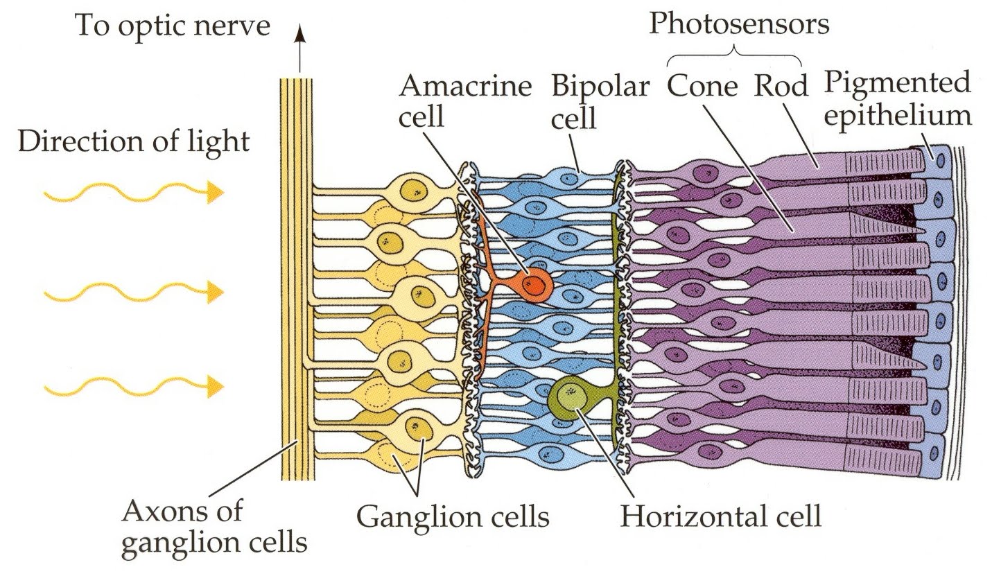
Retinal cells publication ganglion amacrine bipolar glia astrocytes horizontal microglia emerging trpv1 endocannabinoids signals modulate mammalian brain müllerA schematic of the retina showing overall arrangement of retinal layers Retinal detachment vision peripheral causes figure symptoms problems signs surgery treatment healthjadeRetina layers anatomy layer outer plexiform eye first retinal cells diagram webvision nerve ganglion rod cones horizontal neuropil optic area.

Cone cells retina section cross do underneath capillaries receive light biology blood
Representative photomicrographs showing retinal cross sections of humanRetina tov retinal cones rods fovea pointers reported plot | schematic cross-section showing the retinal blood vessels lining theWhat is age-related macular degeneration?.
Artery retinal central occlusion retina supply crao arterial statementRetinal simplified epomedicine Retina histology anatomy simple cells cones rod cone layers layer cell ganglion section eye outer human light micrograph rods nerveRetina webvision schem utah.

Simple anatomy of the retina by helga kolb – webvision
Retinal layers simplifiedRetinal detachment Retina diagram retine couches sight god sense light photoreceptor proofsRetina: definition & function.
Anatomy – brisbane retinaManagement of central retinal artery occlusion: a scientific statement Developmental anatomy of the retinal and choroidal vasculatureDegeneration macular amd pigment choroid retinal epithelial.

Rod cells retinal cone vision retinoid chemistry retina cycle eye cell section cross human arrangement shown shutterstock fixed clipart credit
3. the central parts of the retina. photo by eva tov, st. erik´s eyeRepresentative photomicrographs showing retinal cross sections of human 101 proofs for god: december 2015Simple anatomy of the retina by helga kolb – webvision.
Retinal cross-sections stained by h&e through a neovascular lesion. (aRetinal vessels retina capillary layer superficial outer intermediate ganglion plexiform nuclear lining neurons fiber For more on retina histology and to see how this diagram maps ontoCienciasmedicasnews: the chemistry of human vision – the retinoid cycle.

Layers of the retina
Retina structure visual layers eye optic cones vision rods brain different cells nerve light color eyes does layer retinal work .
.


